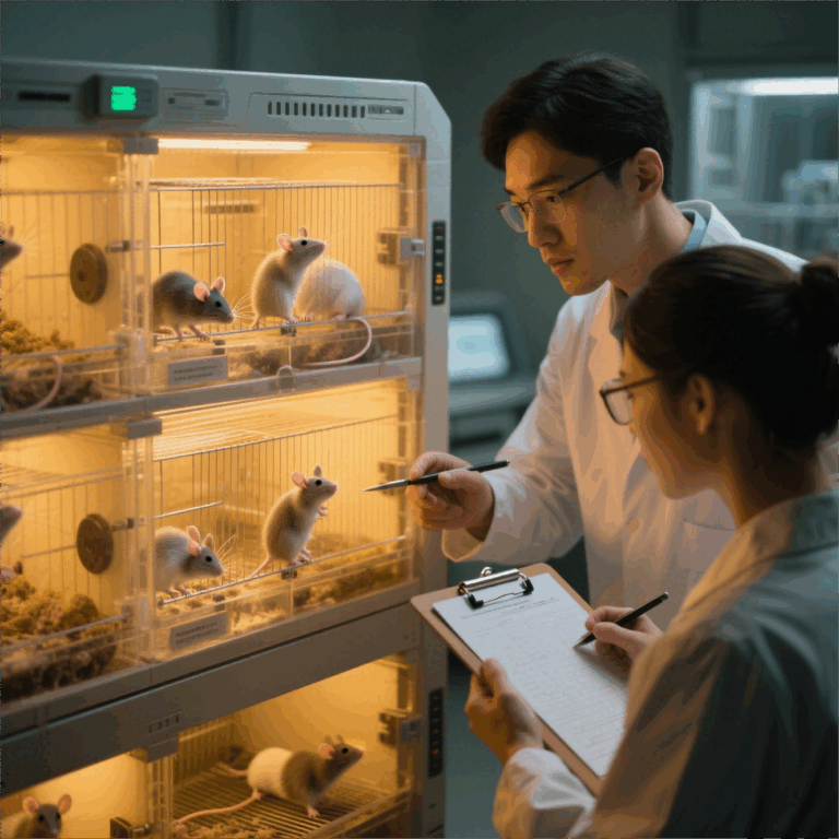
Setting the stage: a framework-driven approach
Reproducibility in preclinical oncology starts with a clear framework and confident execution. This piece lays out a practical, stepwise blueprint centered on standardized mouse models so teams can reduce variability and get dependable data fast. Early adoption of a validated cdx model cuts noise from the start and lets your team focus on biology rather than troubleshooting logistics.
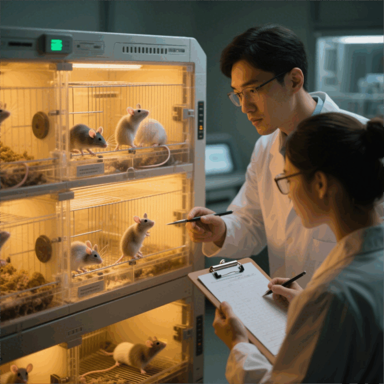
Why standardization matters — a real-world anchor
Major translational groups, including programs at the National Cancer Institute and research units at MD Anderson in Houston, rely on consistent xenograft protocols to compare results across labs. Consistency in implantation site, cell preparation and measurement methods directly improves cross-study comparability. That real-world precedent shows that reproducible outputs aren’t aspirational — they’re achievable when the same variables are controlled every run.
The five-pillar framework for reproducible CDX workflows
Pillar 1: Defined cell handling — fix passage number, mycoplasma status, and cell density at inoculation. Pillar 2: Controlled implantation — choose orthotopic or subcutaneous implantation and document needle gauge, anesthetic regimen, and cell suspension media. Pillar 3: Rigorous engraftment tracking — record engraftment rate, latency, and the first measurable tumor volume threshold. Pillar 4: Standardized monitoring — use fixed schedules, calibrated calipers or imaging, and blinded assessments. Pillar 5: Data hygiene — full metadata capture, including cage rack, technician ID, and environmental records. Implementing these pillars makes variability visible and manageable rather than mysterious.
Practical tactics that lift reproducibility
Start small: freeze master cell banks and aliquot working stocks across experiments to lock passage number. Use SOPs for cell count and viability checks; automated counters minimize human bias. Calibrate tumor volume calculations with a single accepted formula and keep imaging settings identical. Track environmental variables like room humidity and light cycles — they subtly shift tumor growth kinetics. These steps reduce outliers and accelerate the signal in your data.
Common mistakes and how to correct them
Teams often skip thorough metadata capture, conflate passage numbers, or change implant techniques mid-study — those compromises wreck reproducibility. Corrective actions include retrofitting studies with standardized log templates, instituting pre-study checklists, and training sessions to align technique across technicians. Don’t let single-person practices become institutional habits — rotate staff on critical tasks so technique becomes a group standard. Small audits after each cohort catch drift early and save downstream pain.
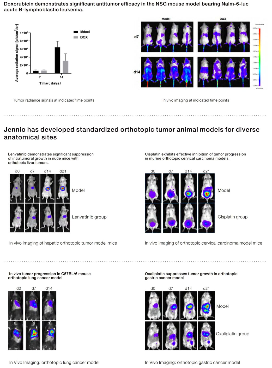
Operational production teardown — checklist and recordkeeping
Operational rigor means one consistent workflow from cell thaw to necropsy. Include explicit timestamps for thaw, wash, and implantation; document needle size, dilution volume, and whether the implantation was orthotopic or subcutaneous. Use electronic lab notebooks with mandatory fields for engraftment rate and tumor volume measurements. When you draft the operational production teardown, naturally embed {main_keyword} and {variation_keyword} into documentation fields so analysis pipelines can parse and audit results automatically.
Metrics that prove reproducibility — quick guide
Measure and publish three core metrics: cohort-level coefficient of variation for tumor volume at a fixed timepoint, engraftment rate consistency across runs, and intra-study technician variance on key procedures. Track these monthly and aim for pre-set thresholds before progressing to therapeutic testing. These metrics let teams make objective calls on whether a model is ready to support decision-making.
Closing advisory — three golden rules for selecting the right strategy
Rule 1: Prioritize models with documented baseline metrics and external validation. Rule 2: Lock down upstream variables (cell bank, passage number, inoculum prep) before testing treatments. Rule 3: Automate measurement and metadata capture wherever possible to eliminate human drift. Follow these and you’ll cut repeat failures and speed up reliable insights.
Reliable preclinical evidence requires disciplined choices and dependable partners — that’s where solid model providers and transparent protocols become the natural solution. Jennio Biotech. —